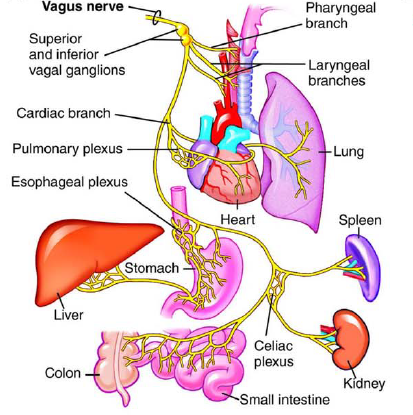
The vagus nerve (VN) is the 10th and most complex cranial nerve that extends from the brainstem to the abdomen and innervates several organs (Martins et al., 2021).
This cranial nerve is primarily cholinergic in nature; however, noncholinergic neurotransmitters also play a role in VN functions. The vagus nerve is considered a protective, complex neuro-endocrine-immune network that modulates the autonomic, cardiovascular, endocrine, respiratory, gastrointestinal, and immune systems (Yuan and Silberstein, 2016a).
The VN consists of afferent (80-90%) and efferent (10-20%) fibers and represents the parasympathetic branch of the autonomic nervous system.
The motor activity of the VN originates from two regions of the brainstem (dorsal motor nucleus and ambiguus nucleus) (Martins et al., 2021).
The afferent fibers end in the area postrema (AP), the spinal nucleus of the trigeminal nerve (SNT), and the nucleus of the solitary tract (NST) (Yuan and Silberstein, 2016a).
The different fibers from the dorsal motor nucleus and the afferent fibers from the solitary tract nucleus form the dorsal vagal complex. This complex controls the subdiaphragmatic organs and conducts the somatic, gustatory, and visceral sensations (Martins et al., 2021).
Efferent fibers from the ambiguus nucleus and afferent fibers that end in the nuclei of origin of the facial and trigeminal nerves control the supradiaphragmatic visceral organs (heart, bronchi, larynx, pharynx, esophagus, and somatomotor pathway) (Martins et al., 2021).
The vagal afferent fibers convey physiological information from the connected organs to the central nervous system. They sense various stimuli, including pressure, pain, temperature, inflammation, etc. (Yuan and Silberstein, 2016a).
Vagus nerve anatomy
Figure 1: Yuan and Silberstein, 2016a
The vagus nerve contains A-, B-, and C-fibers (Yuan and Silberstein, 2016a).
- A-fibers: The large myelinated fibers carry somatic information, whereas the small myelinated fibers carry visceral information.
- B-fibers: Provide efferent sympathetic and parasympathetic preganglionic innervation.
- C-fibers: These small myelinated fibers carry afferent visceral information.
Structurally, VN fibers emerge from the medulla and enter the jugular foramen. After passing through the ganglia, the nerve divides into five branches (Yuan and Silberstein, 2016a):
- auricular branch
- meningeal branch
- sympathetic branch
- pharyngeal branch
- laryngeal branch
The VN travels along the carotid artery and jugular vein and bifurcates to form the recurrent laryngeal nerve (RLN) which sends branches to the larynx and upper esophagus (Yuan and Silberstein, 2016a).
From the upper esophagus, the VN travels downward to the lower esophagus, heart, lung, and related plexuses, and through the diaphragm to the abdominal organs.